Japanese

Chikayoshi Sumi Laboratory: Bio-Medical Engineering Lab.
-Dept of Information and Communication Sciences, Faculty of Sceince &
technology, Sophia University
-Information Science, Graduate School of Sophia University
-Green Science and Engineering, Graduate School of Sophia University
Links
-Laboratory and lectures
Sumi Labolatory Members (passwd required)
Educations, Researches and Grants, etc.
Lectures
-Within University
Department of Information and Communication Sciences
-Department's homepage
Information Science
-Information Science's homepage
Green Science and Engineering
-Green Science and Engineering's homepage
Faculity of Science & Technology
Graduate Division of Sceince & Technology
-Faculity and Graduate Division's homepage
Home page of Sophia University
Yotsuya Campus Access Guide
-Societies
The Japan Society of Ultrasonics in Medicine・Engineering Fellow
The Institute of Electronics, Information and Communication Engineers・Technical
Committee on Ultrasonics
The Japanese Society for Medical and Biological Engineering
The Japan Sports Oryhopaedic Association
The Japanese Orthopaedic Association
The Japan Society of Applied Physics
The Japanese Society for Non-Destructive Inspection
The Japanese Society for Food Science and Technology
The Japan Society for Food Engineering
The Japan Institute Energy
Japan Society of Energy and Resources
The IEEE Senior Member
Our Research Targets
We are conducting development of low-invasive diagnosis/treatment modalities
for various cancerous diseases and various functional disorders, e.g.,
blood flow, nerve and motor. Suitable utilization of electromagnetic waves
and/or mechanical waves and development of solver about inverse problems
will allow providing us innovative clinical modalities and the health devices.
Our current research targets are briefly as follows:
1) Diagnosis: Elasticity Reconstruction, Elastography
Ultrasound imaging,
OCT,
Infrared light imaging,
NMR imaging,
Measurement and analysis of various self-emanating signals,
Deep learning about medical image and data, automatic diagnosis,
etc.
2) Cure: Hyperthermia,
Artificial organs,
Drug deliverly systems, etc.
We work in transdisciplinary fields, contributing to remote sensing and measurement system engineering for cellular engineering, food engineering, and nondestructive examination; ergonomics such as deep-learning computational clones including the linkage of in silico, sensing, robotic devices, and biology; and environmental, energy and SDG reserach, among others.
Samples of Ultrasonic Doppler-Measurement-Based Elasticity Images, i.e.,
Shear Modulus and Strain Images
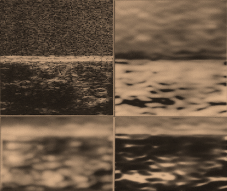
Sample 1: Breast scirrhous carcinoma tissue shear modulus images of a 48.0 mm x 44.6
mm ROI spreading from 17.1 to 65.1 mm in the axial direction. The reference
material (thickness, 40.0 mm) covering the volunteer skin surface has a
shear modulus of 1.2 x 10⁶ N/m2.
B-mode image under pre-compression (upper left, Nominal frequency, 7.5
MHz), and reconstructed absolute shear modulus images in the log gray scale
(upper right, Inverse shear moduli; lower left, shear moduli; lower right,
shear moduli with a high resolution). The highest value: 2.35 x 10⁷N/m2. Upper right (DR: 65.4 dB, the lowest value: 1.26 x 10⁴ N/m2, bright region relatively low shear modulus value), lower left and lower
right (DR: 54.9 dB, the lowest value 4.21 x 10⁴ N/m2, bright region high shear modulus).
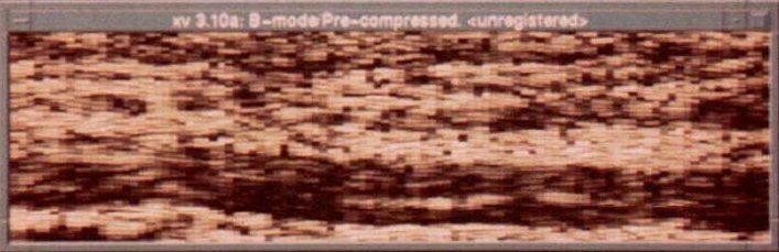
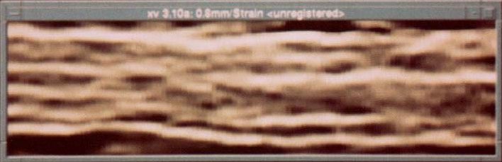
Sample 2: Images obtained on the fresh in vitro pork rib (ROI size, 20.0 mm x 72.8
mm).
(Left) Conventional B-mode image (Nominal frequency, 3.5 MHz).
(Right) Strain image in a log gray scale with a dynamic range of 14.4 dB, in
which the bright region indicates that the region is relatively soft and
vice versa. As shown in this image, the multiple-layer
structure of fat and muscle is favorably visible.
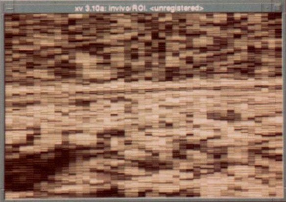
Sample 3: Images obtained on the in vivo breast tissue (healthy 37-years-old, ROI
size, 19.9 mm x 30.0 mm).
(Left) B-mode image (Nominal frequency, 3.5 MHz).
(Right) Strain image in a log gray scale with a dynamic range of 11.1 dB. The reference material attached on her skin surface has a shear modulus of 2.8 x 10⁵ N/m2, and this image exhibits the spatial variation of shear modulus from 4.1
x 104 N/m2 to 5.2 x 10⁵ N/m2. Also in this elasticity image, the fat seems to be favorably visible
(lower left area). Namely, it should be rather soft. Interestingly in this
image, the mammary glands being stiffer seems to be visualized below the
skin surface and also at the lower right area though it is difficult to
detect them from the B-mode image.
Copyright © 1998 Sumi Laboratory, Sophia University. All rights reserved.